
“The foetus was moving, not really sucking its thumb, but it was moving and you could see everything—heartbeats and umbilical cord and so on. It was extremely beautiful, really beautiful!” – Lennart Nilsson
We cannot deny that the word GOOD NEWS has essentially come to represent the coming of a baby. A newborn always brings a smile to the faces of people. It calls for celebration. Celebration for completing the journey of 9 months beautifully and successfully.
These 9 months are quite rough but memorable for the mother but what makes it intriguing is that it is a place we have all been at, it is our origin, but we are completely unfamiliar to it. It is a mysterious biological seat, doing the greatest magic of earth, that is creating a human. But time and now medical scientists have researched to curb this curiosity.
What if a photographer could also do that?
In the year of 1965, Life Magazine published a 16-page story about human reproduction that was specifically extreme macro photographs of how microscopic fertilization happens in the uterus, how a zygote is formed and how the fetus develops inside the womb of a mother. Thus, a different but astonishing form of photography was born: embryo photography.
Swedish photographer Lennart Nilsson spent 12 years taking very close and detailed pictures of the foetus developing in the womb. During the mid-1950s he began experimenting with new photographic techniques to make extreme close-up photographs.
These methods were combined with very thin endoscopes that became available in the mid-1960s, and this enabled him to make cutting-edge photographs of living human blood vessels and body cavities.
He achieved international fame in 1965, with his famous 18 weeks old foetus photograph.
This is something that you’ve never seen before. So, before we dive in, it is to be acknowledged these are actual photographs and not medical scans that are used during pregnancy.
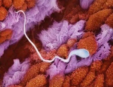
- These are the finger-like projections of the fallopian tube, the tube where the sperm and the egg meet.

Photo by Lennart Nilsson. - This is the sperm gliding in the fallopian tube in search of an egg.
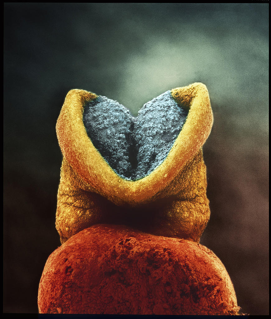
- Oh! The sperm finally finds its soulmate, the EGG
- But it’s not so easy, they won’t have a happily ever after unless the competitors are gone which are about 40-150 million of sperm cells.
- Only one can become the winner, there are no runner-ups in this competition.
- So the winner’s sperm is hailed deep inside the egg and it merges with the egg. This is exactly becoming one
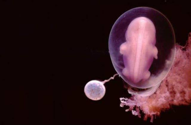
- The outcome of this merging is the zygote, which in 8 days has attached to the wall of the uterus.
Photo by Lennart Nilsson. - Now, this zygote is going to form a baby. So, you can see the brain starting to develop.
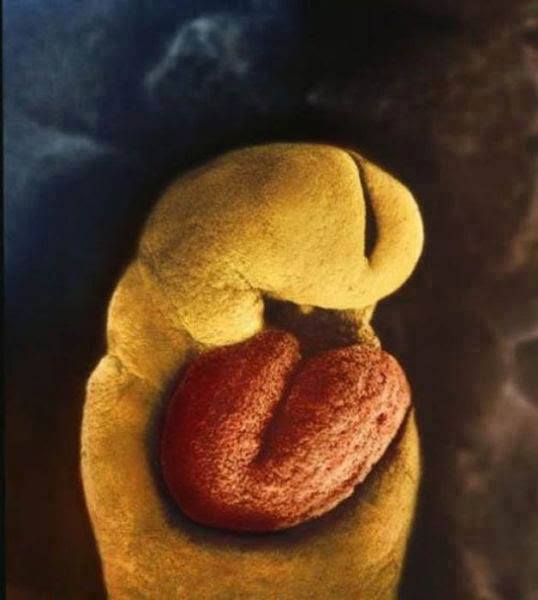
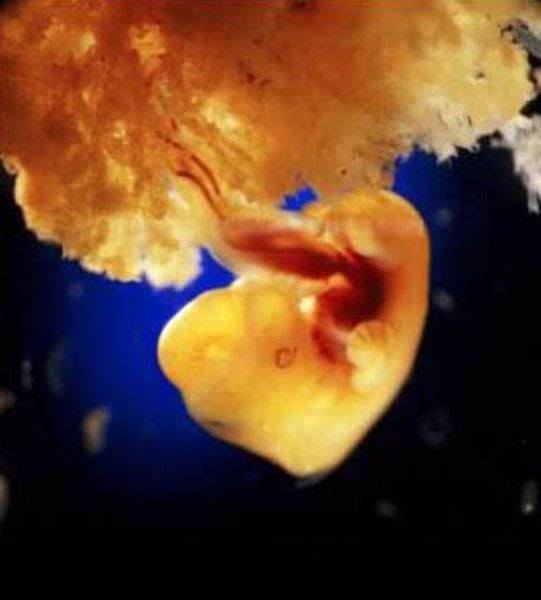
Photo by Lennart Nilsson. - The embryo is a month old; the heart has formed by now and it’s beating. No bones are formed.
- This is what a four-week-old human foetus looks like
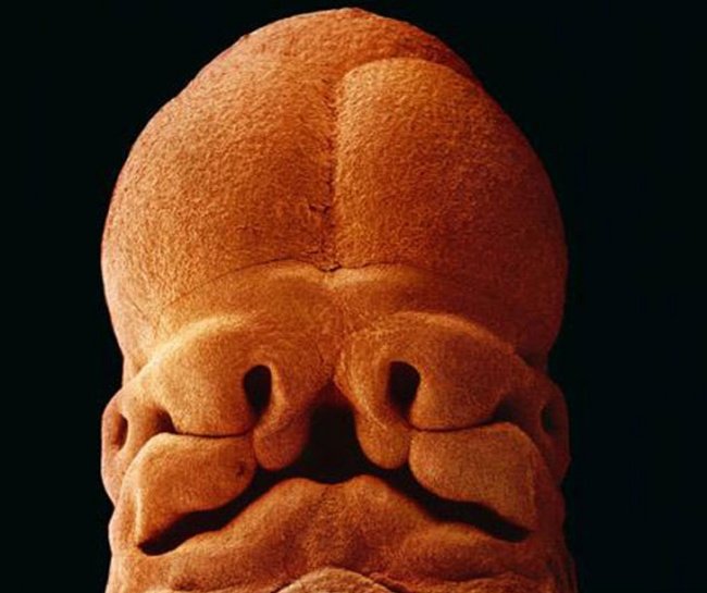
Photo by Lennart Nilsson . - After 5 weeks, this is the face in making. It has an undistinguishable eye, nose, and mouth.
Photo by Lennart Nilsson. - After 40 days the placenta is developed and clearly visible. This is the organ that connects the embryo to the uterine wall. It allows nutrient uptake, waste elimination and gas exchange via the mother’s blood supply.
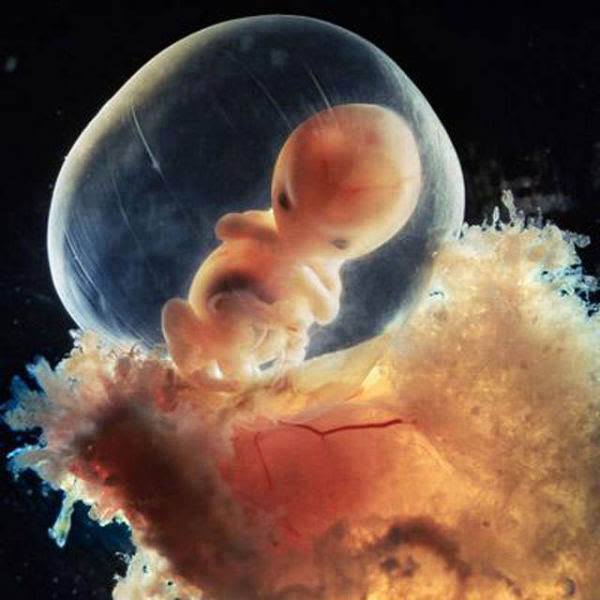
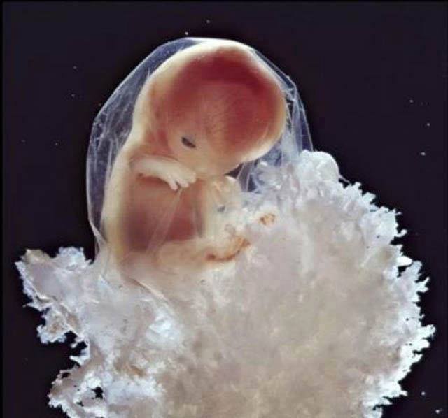
Photo by Lennart Nilsson. - This is an 8-week embryo that is well protected in the fetal sac.
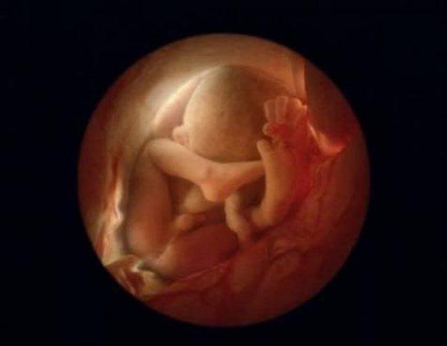
Photo by Lennart Nilsson. - The embryo after 16 weeks
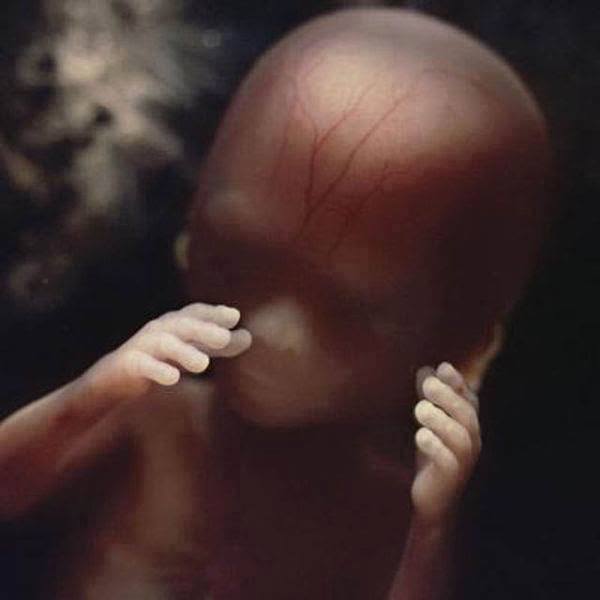
Photo by Lennart Nilsson. - The embryo uses its hand to explore its own body and the surrounding environment
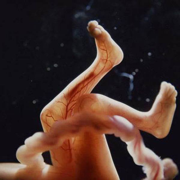
Photo by Lennart Nilsson. - The network of blood supply is visible from the skin because the skeleton has been formed by cartilage which will develop into bones in the later stages of pregnancy. Also, you can see the rope like an umbilical cord.
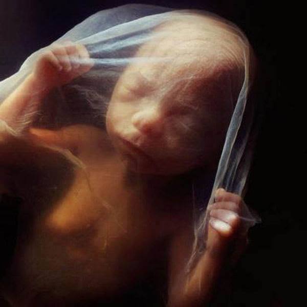
Photo by Lennart Nilsson. - The famous 18-week fetus. It can sense sounds from the outside world
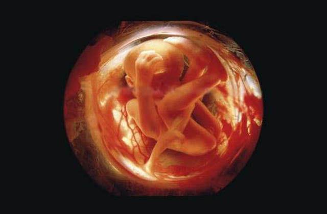
Photo by Lennart Nilsson. - After 26 weeks, the embryo is curled up inside the womb
Photo by Lennart Nilsson. - The fetus generally turns upside down, after 6 complete months, because it is the easier way out into the world
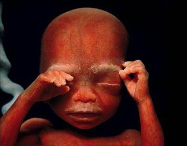
Photo by Lennart Nilsson. - This embryo is about 36 weeks old and it will be able to see the human world in a month.
Nilsson explained in an interview that he had obtained photographs of living foetuses during medical procedures like laparoscopy and amniocentesis and discussed how he had been able to light the inside of the mother’s womb.
He described a shoot that took place during a surgical procedure in Goteborg. He stated, “The fetus was moving, not really sucking its thumb, but it was moving and you could see everything—heartbeats and umbilical cord and so on.
It was extremely beautiful, really beautiful!” Nilsson also acknowledged the fact that he obtained mature human embryos from women’s clinics in Sweden which has been argued by some to be unethical.
But keeping this aside, he received many accolades for photographing the unphotographable.
![]()
